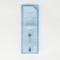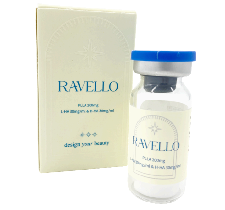

Ravello

$90
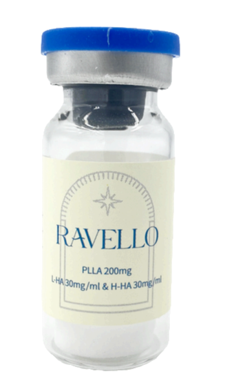
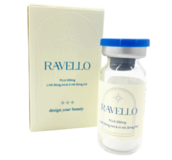
Ravello ®
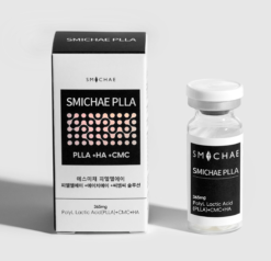
Volume : 200mg / vial (5ml). PLLA 200mg, low molecule 30mg/ml, high molecule 30mg/ml
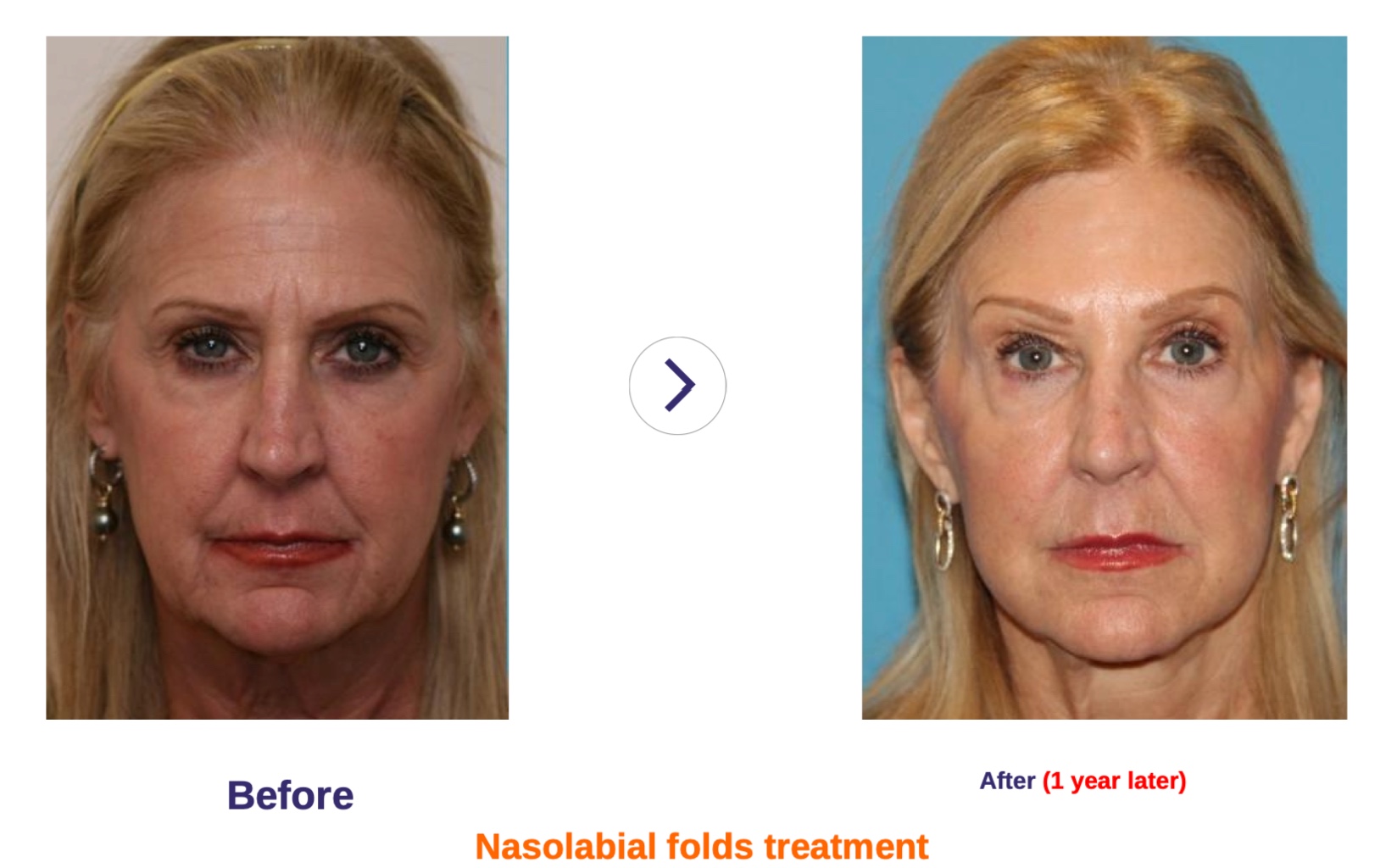
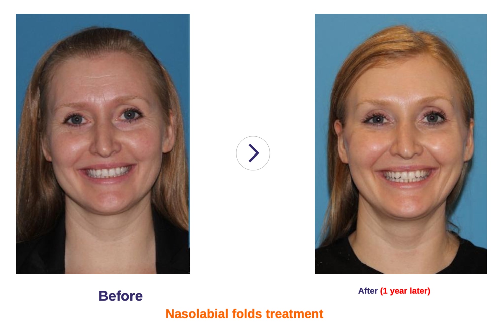
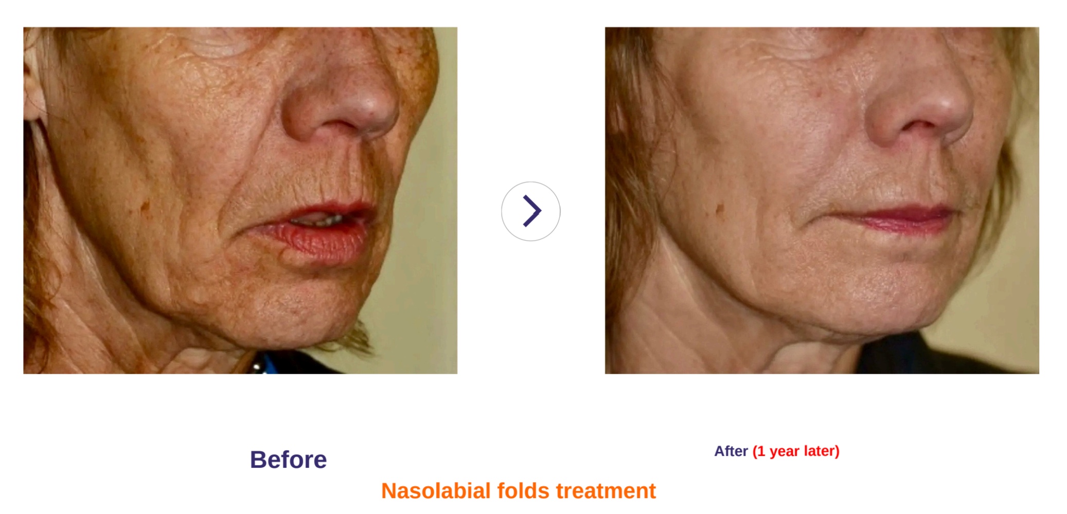
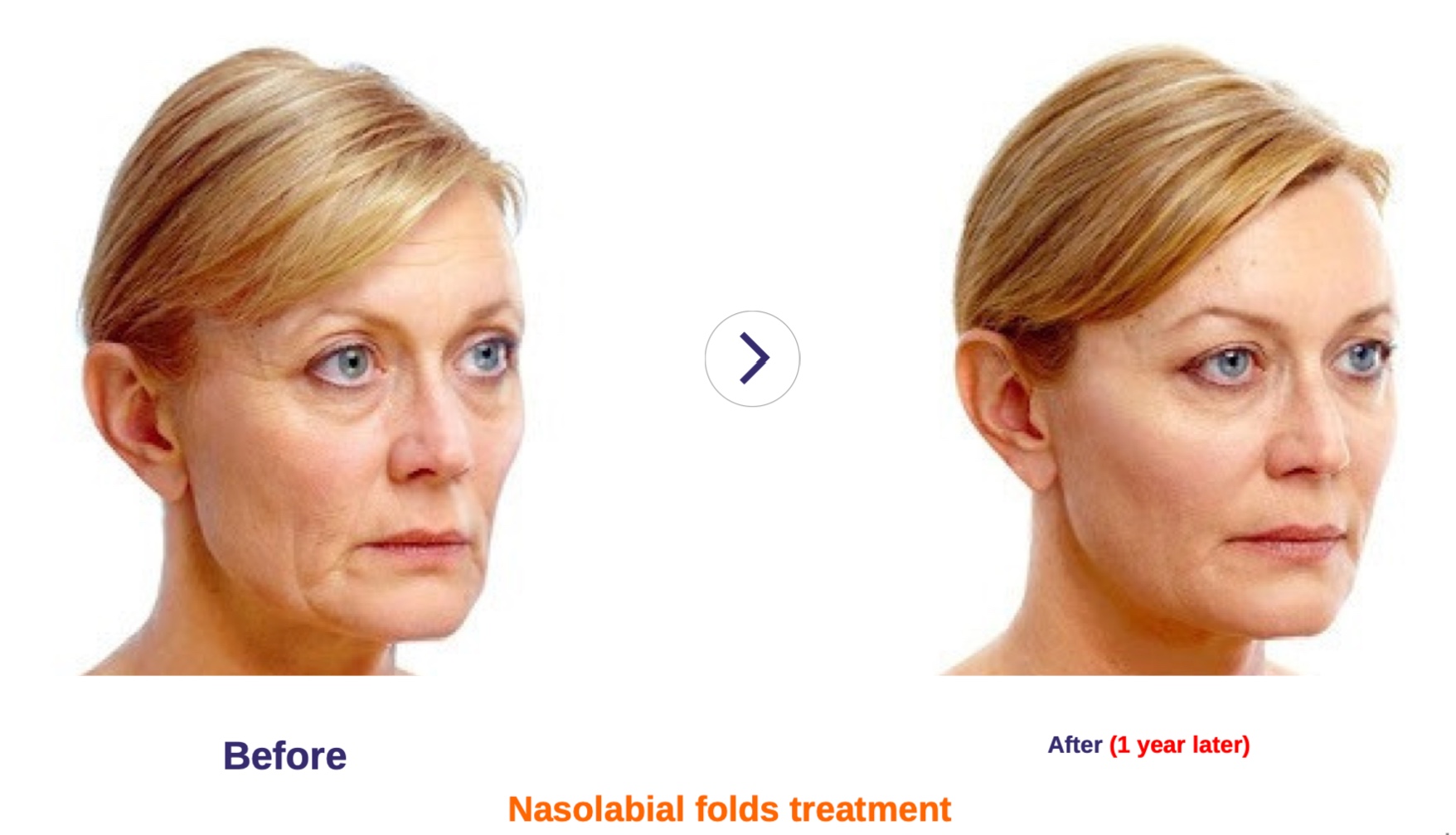
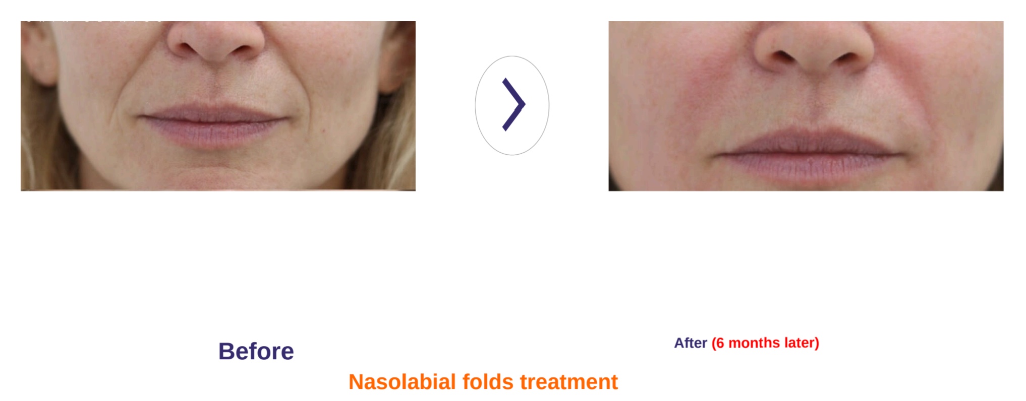
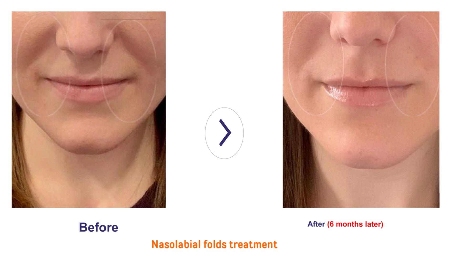
Indication : For treatment of severe facial wrinkles and folds, replacement of volume defects, facial lipoatrophy and improvement official contour
deformities.
RAVELLO PLLA 200mg is a medical device for treating severe facial wrinkles and folds, replacing volume defects and improving facial contour deformities.
As a safe and effective collagen stimulator & regenerator, RAVELLO PLLA helps to restore natural volume for ageing skin-loss collagen. It increases the skin’s elasticity and volume naturally and makes looking young. Depending on the condition of the patients, 2 or 3 times of treatment are recommended for the longevity of volume staying)
Ravello®
Injectable Poly-L-Lactic Acid Powder/HA
PLLA 200mg, Low molecule 30 mg/ml, High molecule 30 mg/ml

WHAT IS Ravello® ?
 Ravello® is a collagen and elastin stimulator made of PLLA and HA which is confirmed by FDA as medical material.
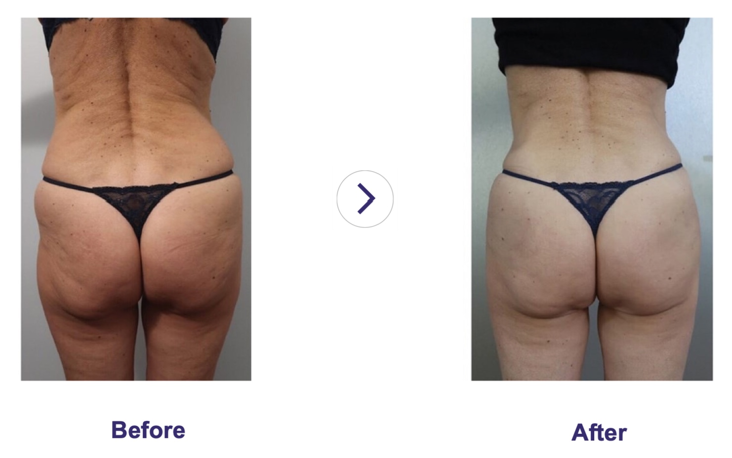
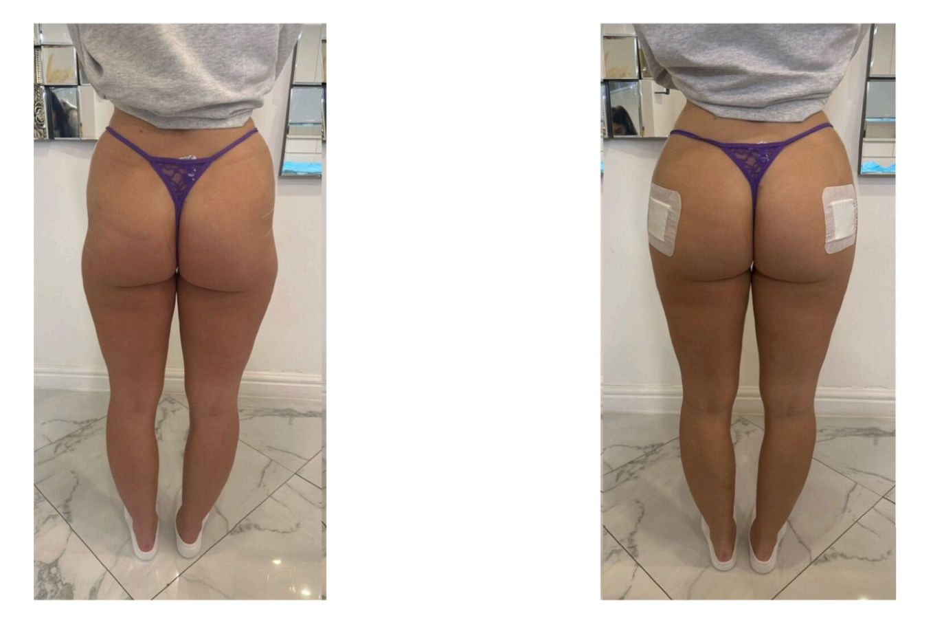
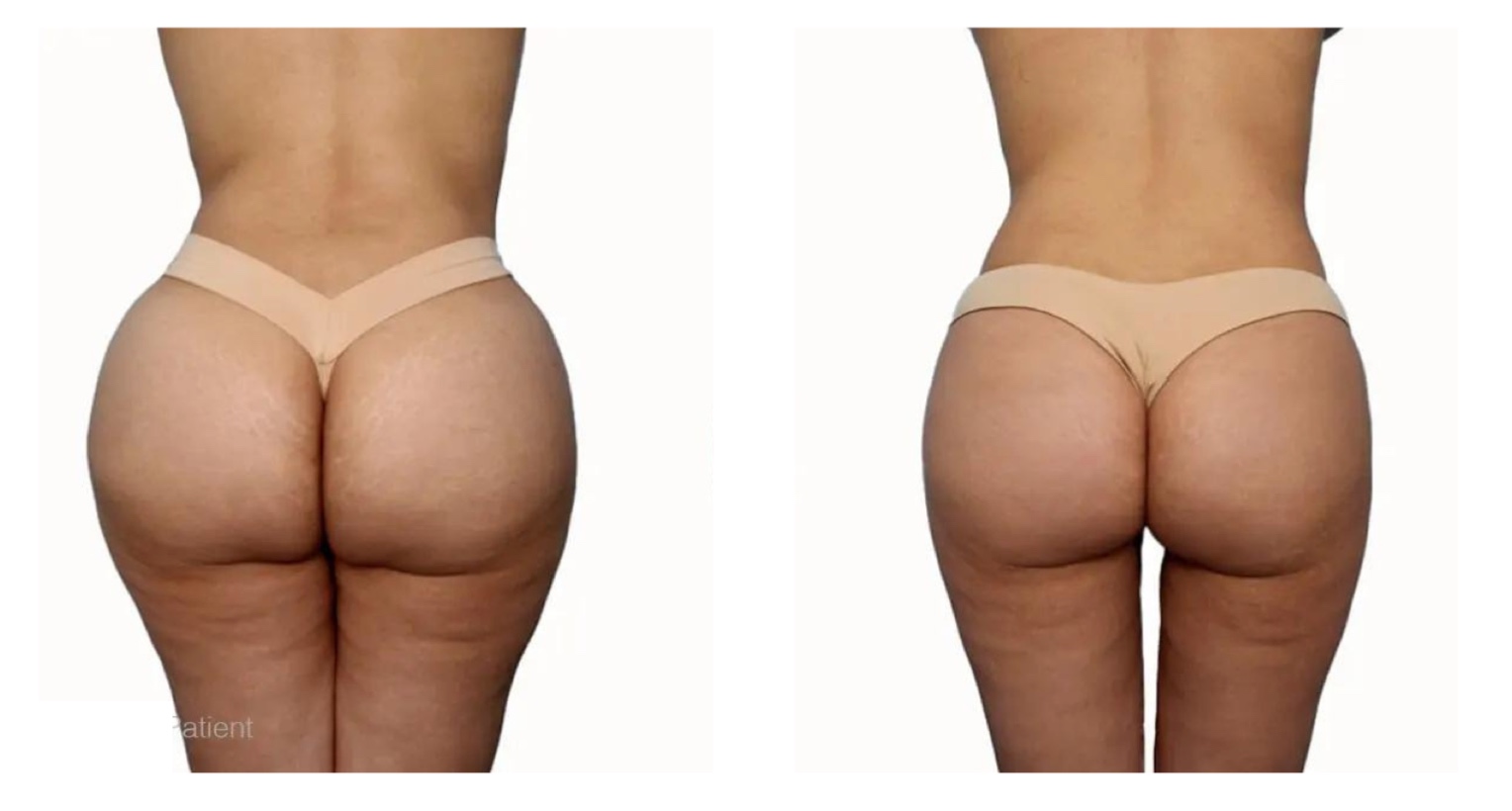
Injected Ravello ® stimulates and regenerates collagen under the skin and restores the skin’s elasticity and volume naturally for facial area, breast, hip, penis, vagina, etc. It helps to reduce fine line wrinkles and present youth looking.
Depends on the condition of patients, 2 to 3 times of treatments are recommended to extend longevity of volume staying.
WHY Ravello® ?
Natural volume with longevity
Injected Poly L-Lactic Acid and HA (Ravello ®) restores volume through stimulating collagen in the skin layer. Generated volume with own collagen lasts more than 2 years.
Confirmed by Korean MDFS.
The main ingredient PLLA is biodegradable.
Solution = Ravello ® + WFl(Water For Injection) +
Lidocaine
is being done over 20 years.
The result will be shown gradually.
With less downtime, easy to return to normal life
Less cost than other surgical treatments
Improvement of wrinkle through lost collagen
regeneration
Feature for Ravello® vs other Type Filler
HA Filler VS Ravello ® VS Fat Graft
Ravello ® doesn’t require retouching for every 6 months but lasts over 2 years after 3 times of initial treatment.
After Ravello ® treatment, patients can able to ordinary daily life.

Ravello ® is a non-surgical treatment and recovery time is shorter than fat graft due to formation of biodegradable scaffold and generation of collagensis, do not need to worry about engraftment of autologous fat.
- In general, patients may experience some degree of swelling because of the injection treatment itself. Because of this swelling, the injection sites may appear to be completely corrected. immediately (within 30 minutes) after injections.
- Patients should be informed that the swelling after the injection will be resolved within hours or days after injection, and that the injection sites will result in the original appearance before the injection treatment.
The correlation between Collagen & Skin aging
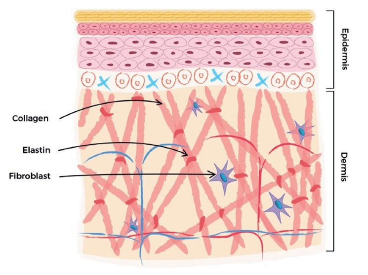
Fig 1. The production of collagen fibres in the dermis.
The fibroblast secretes the procollagen fibre into the extracellular matrix, where they form larger collagen bundles. Elastin is also secreted and assembled into the collagen-based macromolecular structure. (By permission of MINERVA Research Labs Ltd – London)
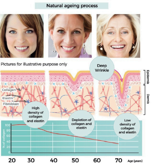
Fig 2. Collagen content of skin is maximal between the 2nd and 3rd decade, after which there is a slow depletion and loss of collagen (and associated ECM components such as elastin and GAGs). The loss of collagen is clearly correlated with changes to appearance attributes which are typically referred to as fines lines and wrinkles. (By permission of MINERVA Research Labs Ltd – London)
- The proportion of the collagen types in skin change with age. Young skin is composed of 80% type I collagen and about 15% collagen type Ill. With age, the ability to replenish collagen naturally decreases by about 1.0%-1.5% per year.
- This decrease in collagen is one of the characteristic hallmarks associated with the appearance of fine lines and deeper wrinkles. [Fig. 2]
- Moreover, deep inside in the dermis, fibrillar collagens, elastin fibers and hyaluronic acid, which are the major components of the extracellular matrix, undergo distinct structural and functional changes.
- The loss of collagen is clearly correlated with changes to appearance attributes which are typically referred to as fine lines and wrinkles.
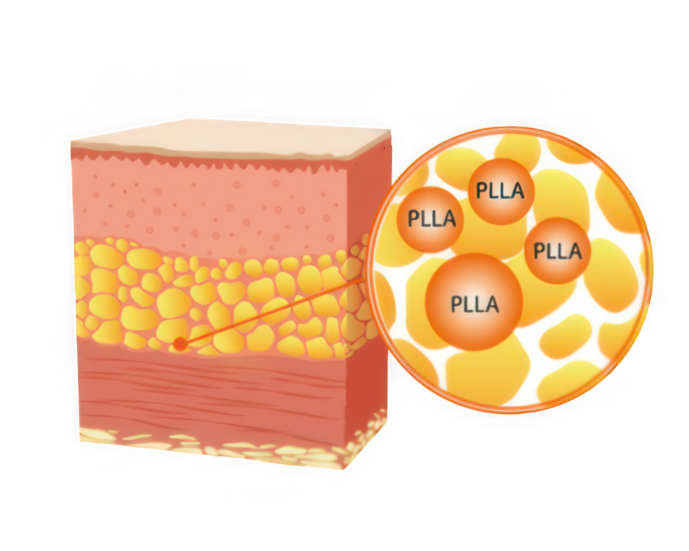
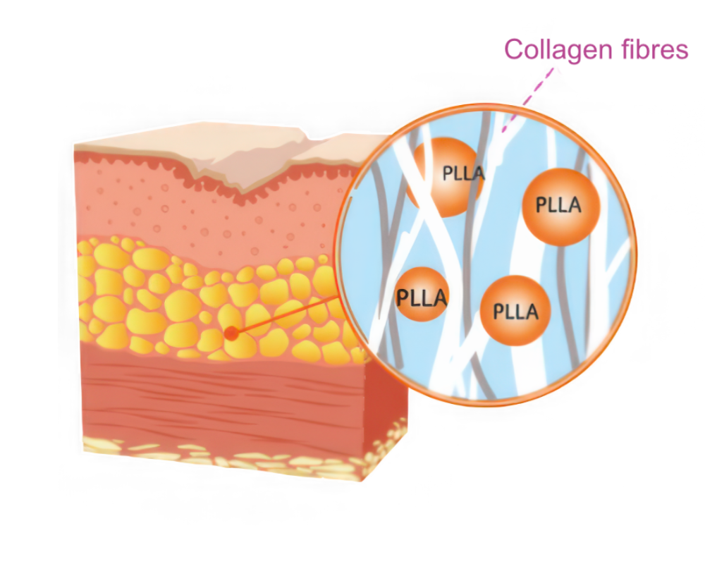
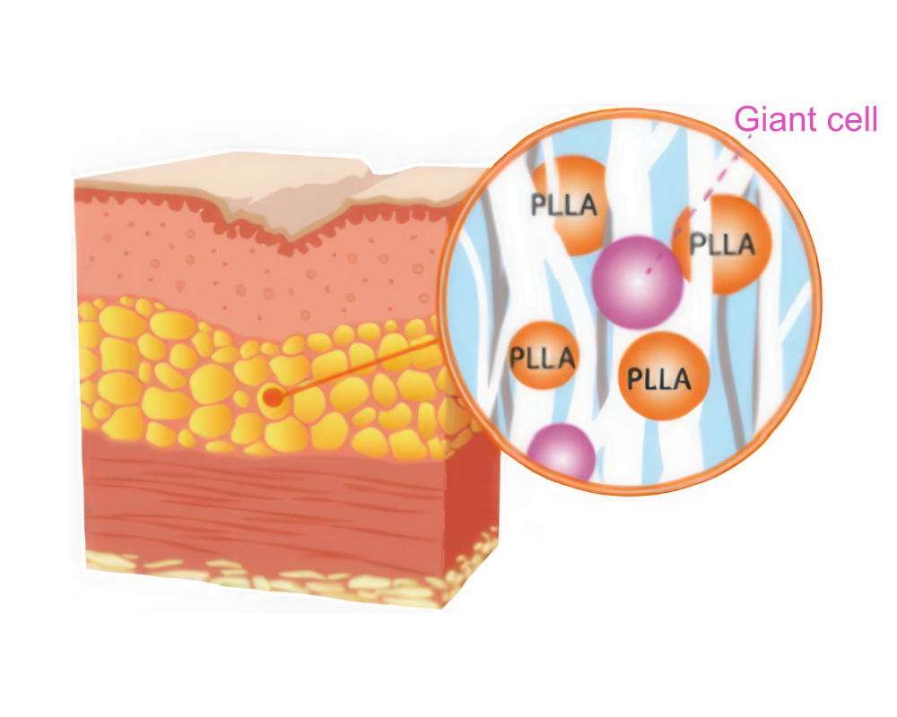
HOW Ravello® WORKS
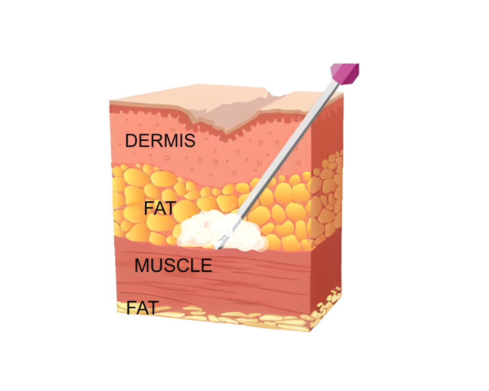
Inject diluted Ravello ® into the subcutaneous layer of the skin.
Firstly wrinkle improvement can be shown with WFl (Water for injection) and diluted Ravello ®
Within some days, after absorbing water, only Ravello ® particles left
New cells are gathered around PLLA & HA and increasing fibroblasts.
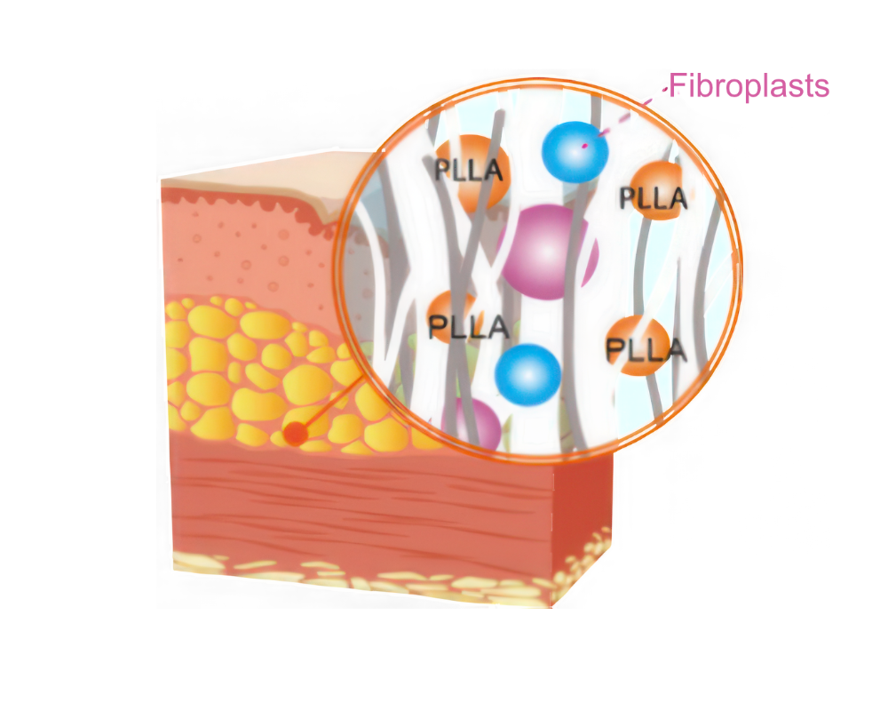
Increased fibroblasts stimulate collagen regeneration and improve skin folds.
Regenerated collagen lasts for 2 years and Ravello ® particles are biodegraded.
Rebuild lost Collagen, Collagen Stimulator as Skin Booster
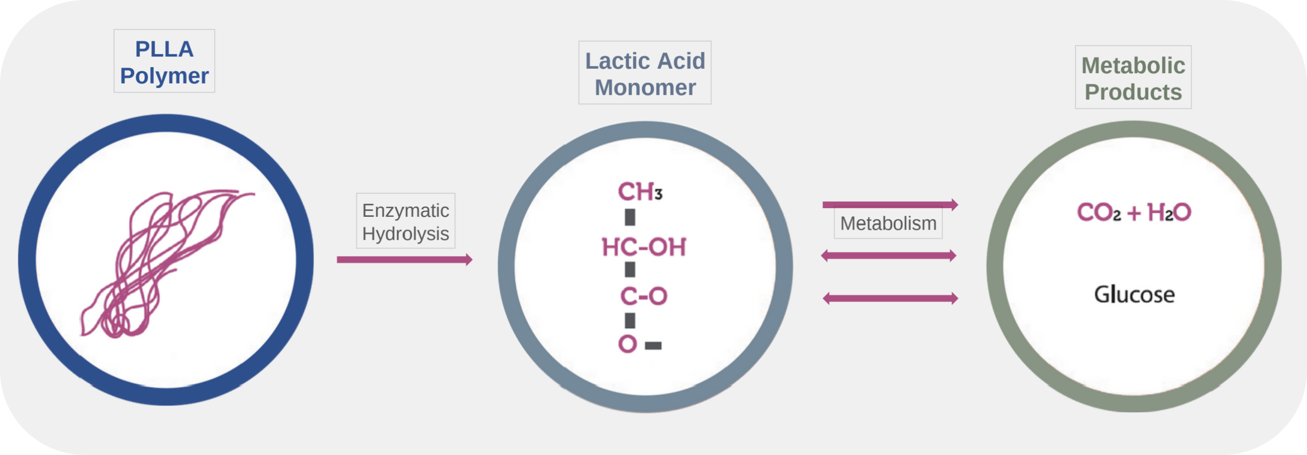
- PLLA(Poly-L-Lactic Acid) is a biodegradalbe raw material approved by the U.S. FDA.
- After injection, PLLA(Poly-L-Lactic Acid) & HA stays for certain time, then it is metabolized and decomposed into lactic acid, H₂O, CO₂ and glucose.
- PLLA(Poly-L-Lactic Acid) is synthetic polymer of lactic acid, the substance that exists naturally in human body.
- Ravello ® consists of a microporous PLLA and HA.
Safe Collagen stimulator PLLA & HA
Ravello ® has reduced the possibility of side effects comparing to other companies.
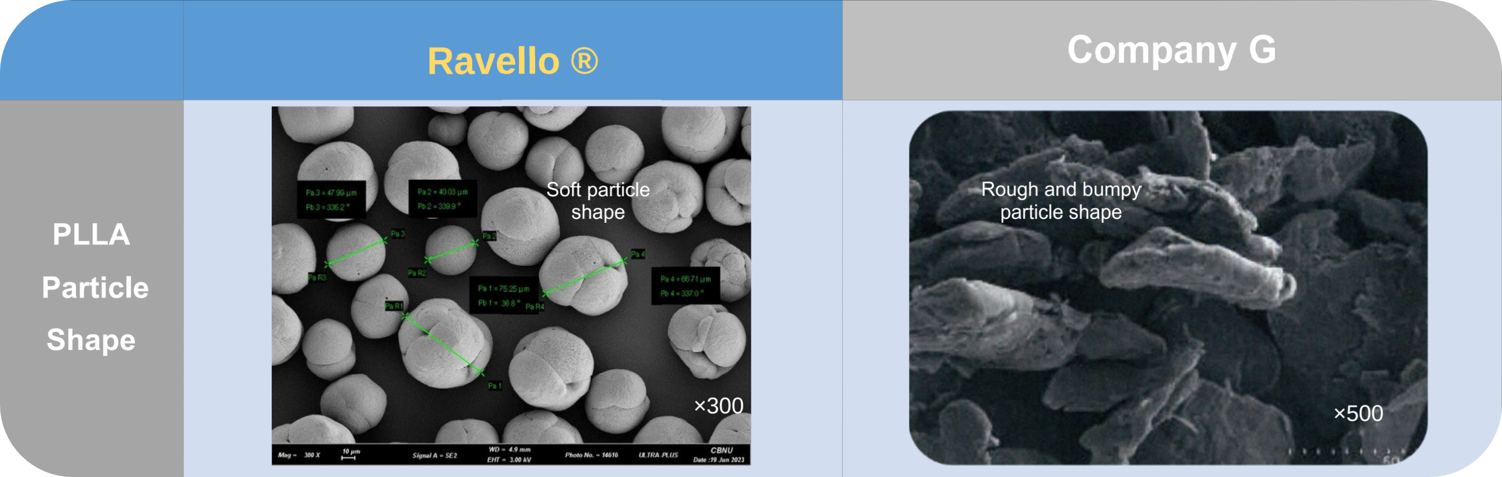
Ravello ® PLLA is a microporous PLLA sphere type. The shape of Ravello ® is soft and round, which can help reduce side effects such as nodules.
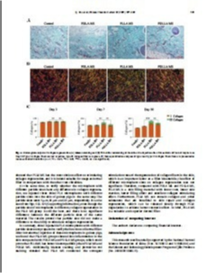
Histopathological and immunofluorescence assessments of inflammatory response
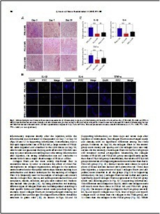
Hematoxylin and eosin (H&E) staining can be used to observe the local inflammatory response and determine the biocompatibility of PLA microsphere. In Fig. 6A, the results showed that no obvious histopathological damage or inflammatory reaction was detected three days after the treatment of PLLA MS, indicating excellent biocompatibility.
As comparison, the inflammatory cells in the PDLA MS group were significantly more than those in the PDLLA MS and PLLA MS group, it proved that the inflammatory response in the skin injected by PDLA MS was stronger than that in PDLLA MS and PLLA MS group.
The expression level of IL-1β, IL-6 and TNF-α were evaluated by confocal laser scanning microscope (CLSM). As shown in Fig. 6B, on Day 3, the fluorescence signal of FITC in PDLA MS group was obviously strong, which showed the level of inflammatory response in tissues surrounding of the injected site with PDLA MS was significantly stronger than the level of inflammatory response which injected with PLLA MS or PDLLA MS.
The semi-quantitative histograms in Fig. 6C showed that the expression levels of IL-1β, IL-6 and TNF-α around the injected site induced by PDLA MS were 3.2, 2.5 and 2.0 times higher than those of inflammatory factors induced by PLLA MS, and 2.3, 1.9 and 1.4 times higher than those induced by PDLLA MS, respectively.
This finding was in consistence with the results in H&E staining, confirming that PLLA MS has good biocompatibility and thus is more suitable as dermal filler (Skin Booster or Collagen Stimulator).
[Reference] Chinese Chemical Letters 32 (2021) 577-582
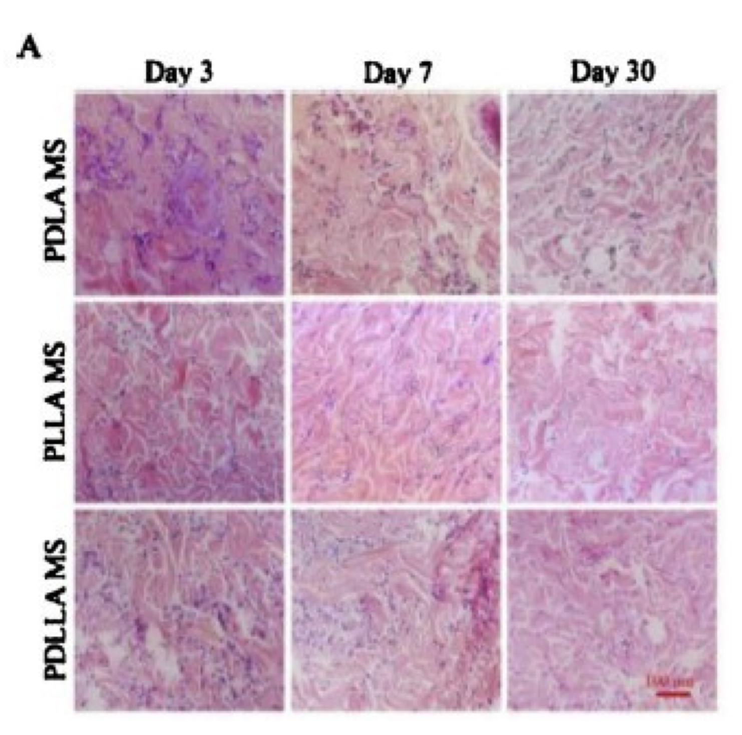
(Fig. 6A) H&E staining of the skin at the injection site of PDLA MS, PLLA MS, and PDLLA MS.
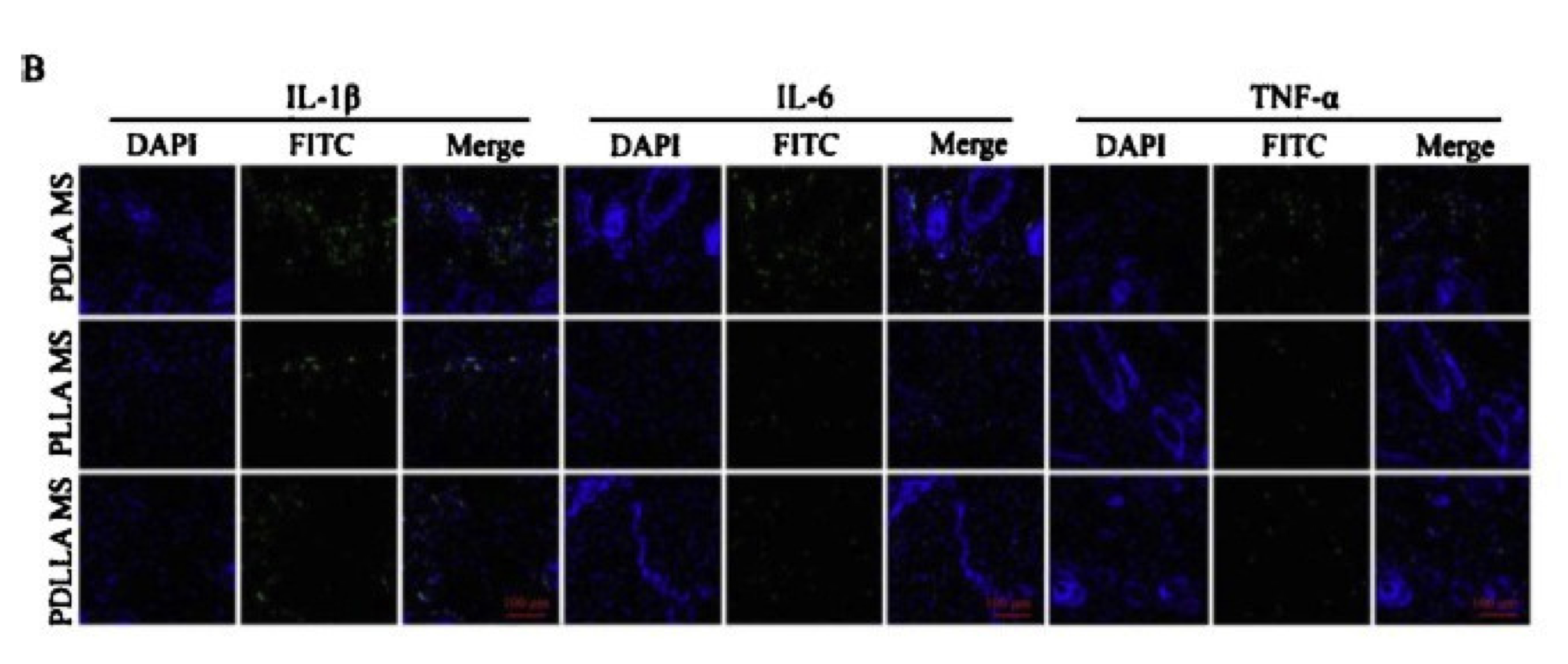
(Fig. 6B) Immunofluorescence determination of skin inflammatory factors on Day 3 after microsphere injection. Green fluorescence showed FITC labeled inflammatory factors and blue fluorescence showed nuclei.
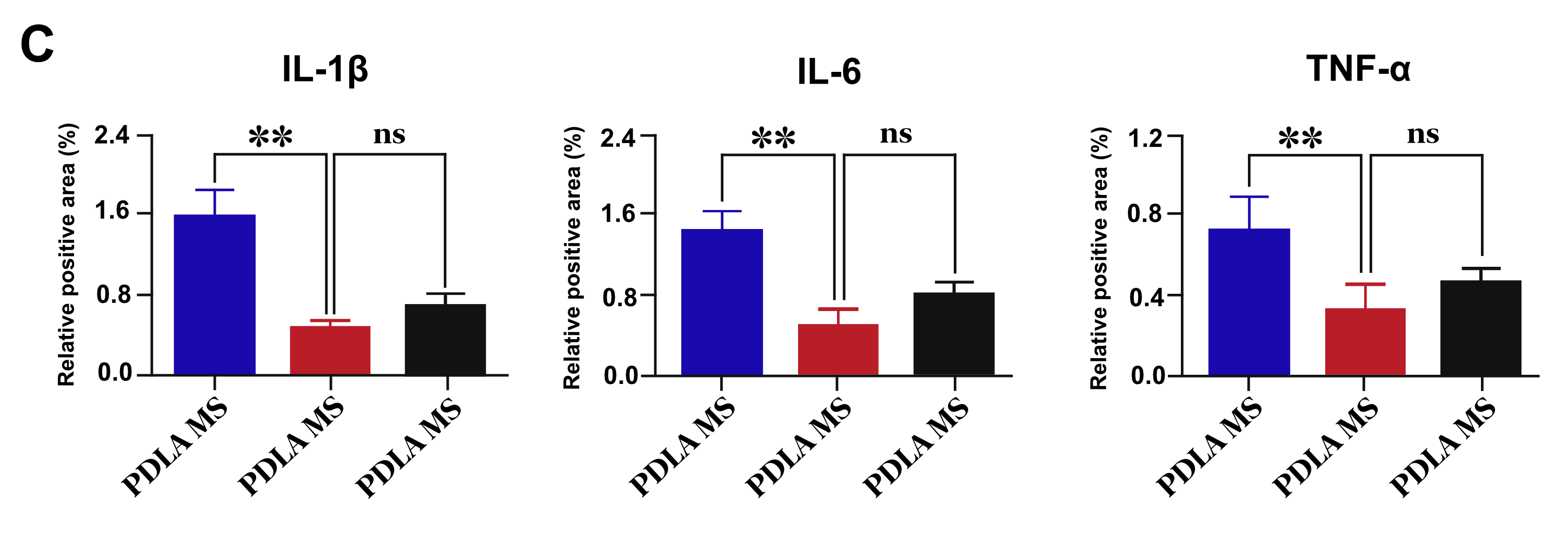
(Fig. 6C) Semi-quantitative analyses of IL-1β, IL-6 and TNF-α.
Data are presented as mean standard deviation (n = 3; *P < 0.05, **P < 0.01, ***P < 0.001, ns: not significant).
Superior Collagen Stimulator PLLA – Histrological Analysis of Collagen Regeneration
[Reference] Chinese Chemical Letters 32 (2021) 577-582
The content of type I collagen in the PLLA group was still 84.7%, and type III collagen was 15.1% on 30 day.
Total of type I and type III collagen in the PLLA group were 1.4 times than the collagen in the PDLA group, and 1.1 times than the collagen in the PDLLA group (Fig. 7C). Which showed that PLLA MS has the most obvious effect on stimulating collagen regeneration, and it is indeed suitable for usage as dermal filler in comparison with the other two chiralities.
Masson staining and picrosirius red staining revealed that PLLA MS conducted the strongest stimulation toward the regeneration of collagen fibers in the skin, which is an important factor as a filler.
Therefore, compared with PDLA MS and PDLLA MS, PLLA MS is a skin filling material with lower cost, lower side reaction, better filling effect and excellent collagen stimulating effect. Furthermore,
PLLA MS can contain collagen and other substances that are beneficial to skin repair and collagen regeneration, which can be released slowly through PLLA degradation to achieve better cosmetic effect.
In brief, PLLA MS is a valuable and superior dermal filler (Skin Booster or Collagen Stimulator).
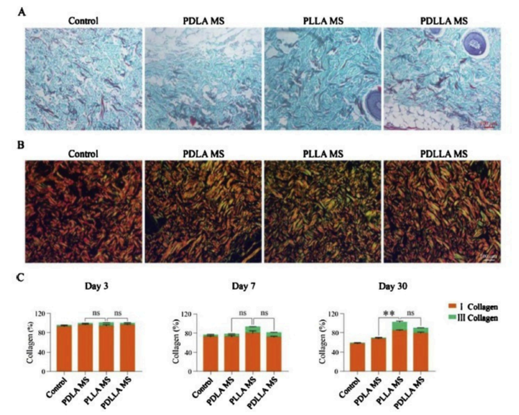
(A) Masson
staincinogll a gen is stained blue, nuclei are stained dark brown, muscle tissue is stained red, and cytoplasm is stained pink
The skin picture at the injection site of three kinds of PLA microsphere on Day 30.
(B) Picrosirius red staining
Picrosirius red (PSR) staining is a commonly used histological technique to visualize collagen in paraffin-embedded tissue sections.
PSR stained collagen appears red in light microscopy.
However it is largely unknown that PSR stained collagen also shows a red fluorescence, whereas live cells have a distinct green autofluorescence
(C) Semi-quantitative analyses of type I and type Ill collagen fibers.
Data are presented as mean A: standard deviation
(n = 3, *P < 0.05, **p < 0.01, ***p < 0.001, ns: not significant)
Type l collagen fibers are red or yellow, type lll collagen fibers are green.
Comparison Of Collagen Regeneration : PLLA Skin Booster vs Control
Masson Trichrome staining
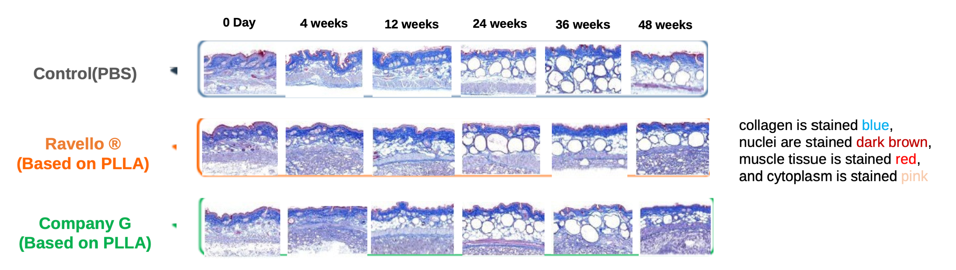
Figure 8. Confirmation of collagen regeneration over time after filler injection – MT stain (X100)
MT staining revealed that PLLA MS conducted the strongest stimulation toward the regeneration of collagen fibers in the skin, which is an important factor as a skin booster compared to control.
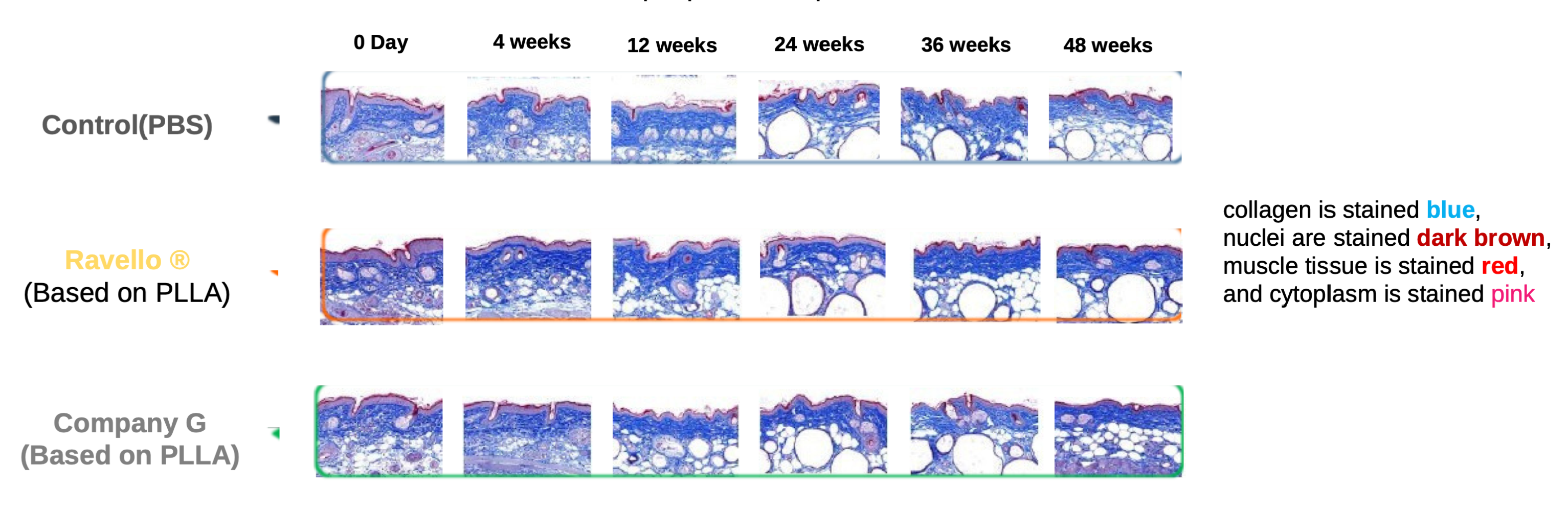
Figure 9. Confirmation of collagen regeneration over time after filler injection – MT stain (X200)
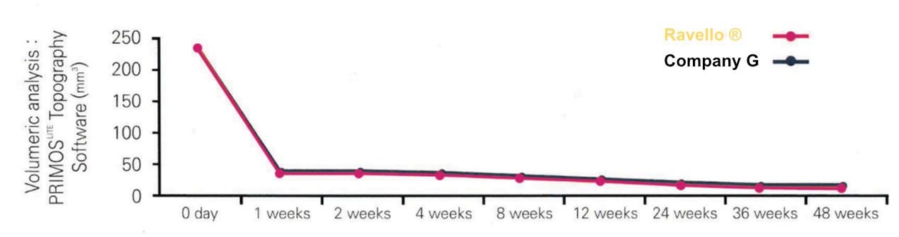
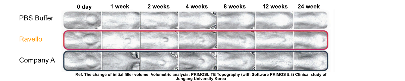
Ravello ® was almost similar to company G’product, a licensed product, as a result of the control test for decomposition.
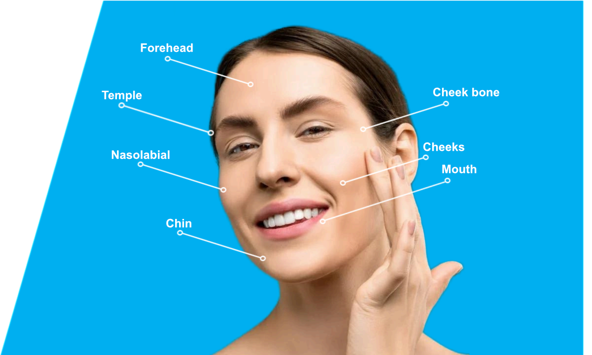
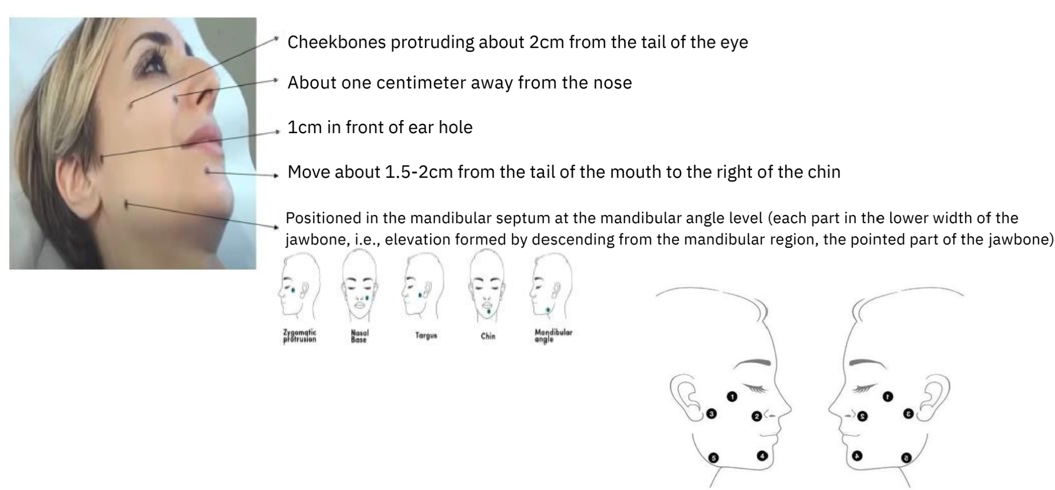
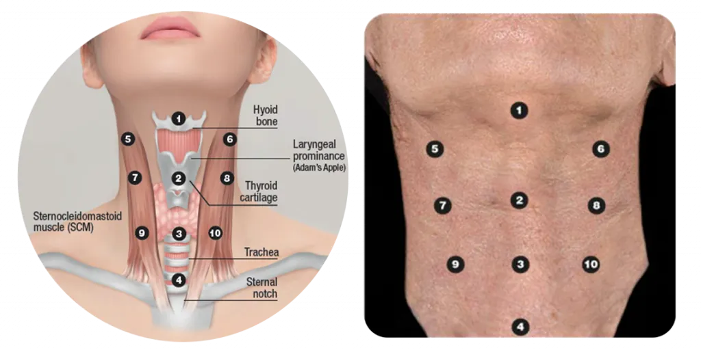
Recommended area for treatment
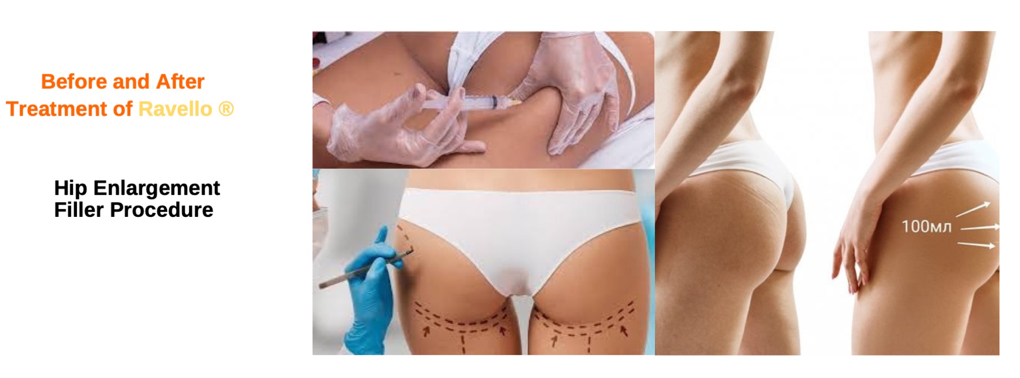
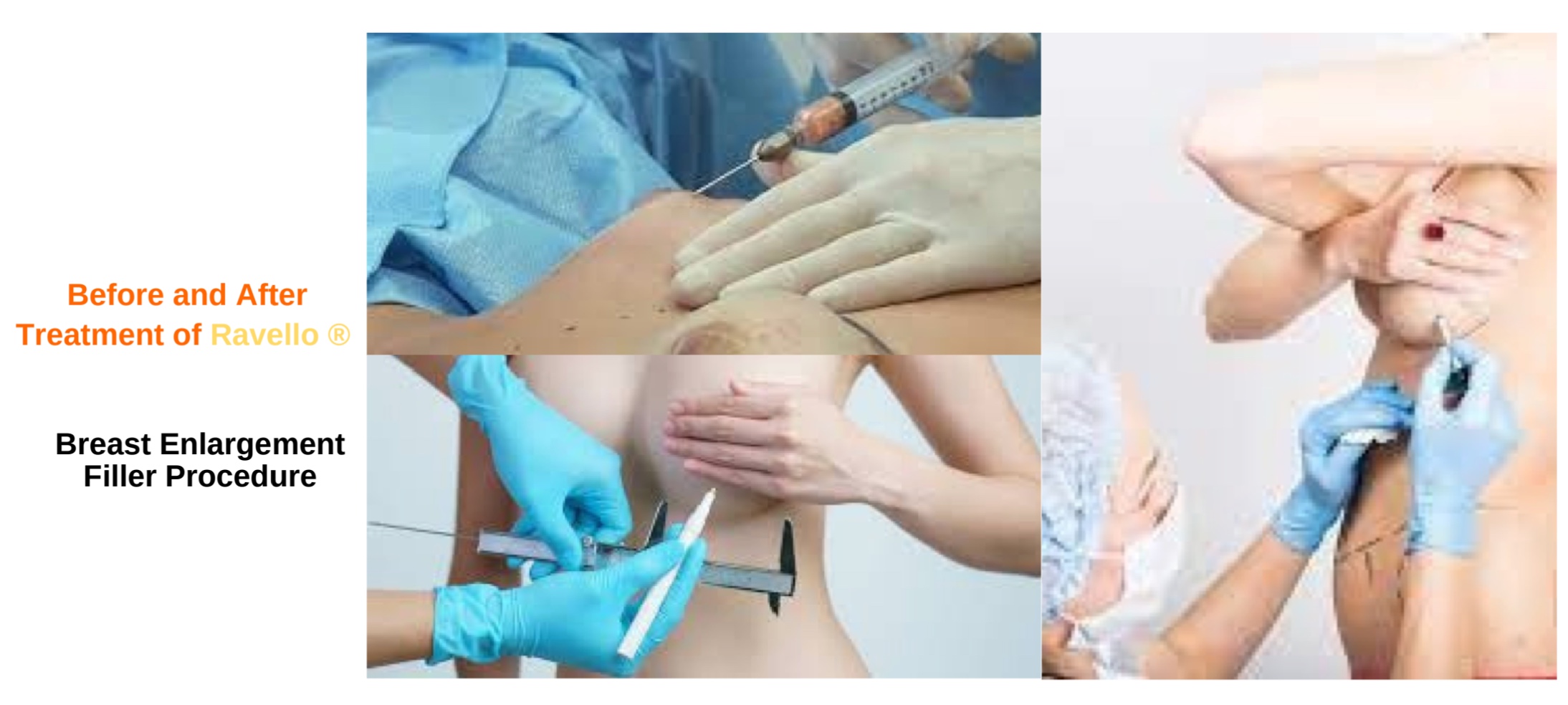
Ravello Treatment Protocol
Based on 260mg/Vial
- Water for injection (sterilized 0.9% saline water) and Lidocane injection
Remove the Ravello aluminum stopper and disinfect the rubber stopper with rubbing alcohol.
Inject 7mL of water for injection and 2mL of 2% Lidocane into Ravello 260mg/Vial. - Hydration After injecting water for injection and lidocane, shake the vial well so that the PLLA is well dispersed in the HA. Let stand for about 1 hour.
- Before the procedure, shake the hydrated Ravello so that the PLLA is completely dispersed in the HA.
Make sure it is well distributed before using it. If too much is transferred to a syringe, PLLA agglomeration will occur inside the syringe. If PLLA aggregation occurs, only PLLA may remain in the syringe and only the HA suspension may be injected.
->Solution: Use a small syringe and perform the procedure using several small syringes. - Procedure Before the procedure, wipe your face with rubbing alcohol on the treatment area and disinfect your face with iodine solution.
The treatment should be performed on the BAP Point, and approximately 0.4 to 0.5 CC per point should be treated in the subcutaneous fat layer. After each treatment point, gently rub to spread the PLLA well. During the procedure, clean the area that bleeds from the needle thoroughly with disinfecting alcohol.
- Consultation and Patient Assessment:
- Assessment of the skin condition, identification of problem areas, and determination of indications for PLLA application.
- Discussion of expected results, potential risks, and possible side effects.
- Preparation for the Procedure:
- Skin cleansing and makeup removal.
- Antiseptic treatment of the injection area.
- If necessary, application of local anesthesia to reduce discomfort.
- Preparation of the Product:
- According to the manufacturer’s recommendations, the product is diluted with sterile water to the required volume.
- It is usually recommended to allow the solution to stand for a certain period to ensure full hydration.
- Marking and Injection Technique:
- Marking the skin in accordance with anatomical landmarks and the correction plan.
- The product can be injected using a cannula or a fine needle into the deep dermis or subcutaneous tissue.
- Even distribution of the product across the treated area to achieve a natural result.
- Post-Procedure Care:
- Gentle massage of the treated areas to evenly distribute the product.
- Recommendations for skin care after the procedure, including avoiding intense physical activity, heat treatments, and facial massage for several days.
- Scheduling a follow-up visit to assess the results and, if necessary, to perform additional procedures.
Important: This protocol is generalized and may vary depending on individual patient characteristics and the preferences of the treating physician. It is strongly recommended to follow the manufacturer’s instructions and to perform the procedure only by a qualified specialist.
Caution: Do not treat the areas specified below:
- Between the eyebrows
- Just above the eyebrows If you want to perform a procedure on the forehead, assuming that the area of the forehead is divided into upper and lower halves, The procedure should be performed as high as possible.
- Lips and just below the lips Assuming that, like the forehead, it is divided into upper and lower halves based on the lower lip and the tip of the chin, perform the treatment as low as possible.
- Under the eyes During the procedure, a 27 Gauge needle or a cannula is used after punching.